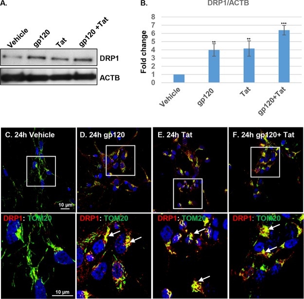
FIG 4.
HIV gp120 and Tat trigger mitochondrial translocation of dynamin-related protein 1 (DRP1), leading to mitochondrial fission and altered mitochondrial dynamics. (A) Western blot analysis of neuronal lysates showing increased DRP1 expression in neurons treated with HIV gp120 and Tat for 24 h. (B) The relative intensities of DRP1 proteins were normalized to the results for beta-actin (ACTB). Each data point was normalized to the results for treatment with vehicle and analyzed using Image J software. Student's t test was performed to test the statistical significance. Data are presented as mean values ± SD (n = 3 independent donors). **, P < 0.01; ***, P < 0.001. (C to F) Confocal laser microscopy analysis shows increased expression and translocation of DRP1 to TOM20-stained fragmented mitochondria. Neurons were immunostained with antibodies specific to DRP1 (red) and TOM20 (green). In the enlarged images of boxed areas, white arrows indicate colocalization (yellow) of DRP1 with TOM20-stained mitochondria. Scale bars, 10 μm.

