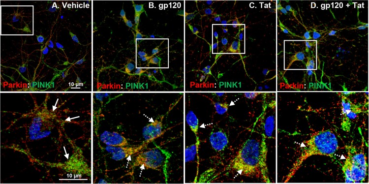
FIG 5.
HIV gp120 and Tat increase PINK1-Parkin immune complex formation and translocation to the damaged mitochondria. (A to D) Confocal laser microscopy analysis shows increased expression, translocation to perinuclear areas, and immune association of PINK1-Parkin complexes (indicated by the dashed white arrows in enlarged images of boxed areas) with the damaged mitochondria in HIV gp120- and Tat-treated neurons. In contrast, vehicle-treated neurons display low, diffuse, cytoplasmic expression of PINK1 and Parkin proteins, as indicated by the solid white arrows. Scale bars, 10 μm.

