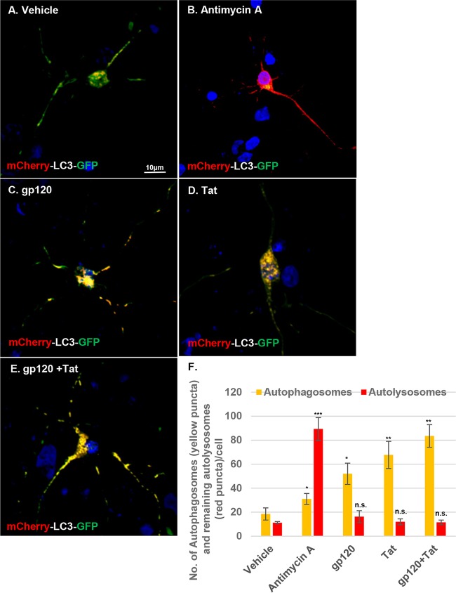
FIG 8.
Treatment with gp120 and Tat induces incomplete autophagy. (A to E) Autophagic flux was monitored using a dual-fluorescence mCherry-EGFP-LC3 vector. HPNs transiently expressing mCherry-EGFP-LC3 plasmid were treated with HIV gp120 and Tat for 24 h. A concentration of 10 μM antimycin A was used as a positive control. Cells were then fixed and analyzed using confocal microscopy. Scale bar, 10 μm. (F) Quantitative analysis of the numbers of autophagosome puncta (yellow) and the remaining autolysosome puncta (red) per cell. Student's t test was performed to test for statistical significance. Data are presented as mean values ± SD (n = 4 independent donors; n ≥ 10 cells per condition). *, P < 0.05; **, P < 0.01; ***, P < 0.001; n.s., not significant.

