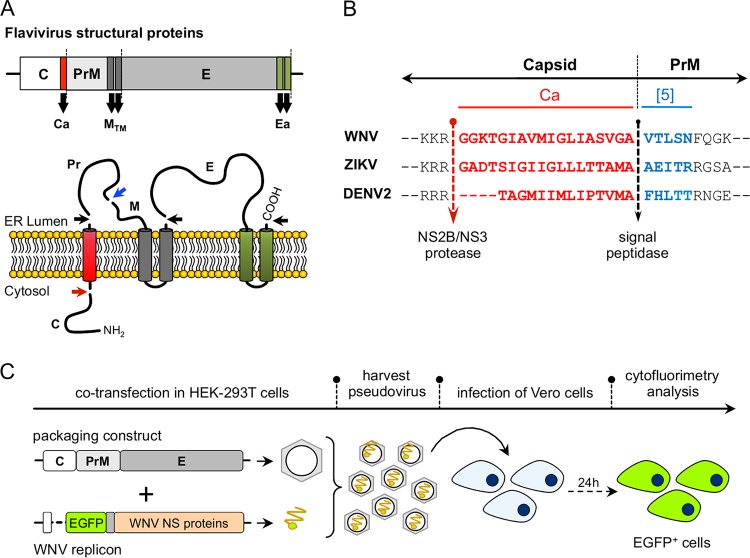
FIG 1.
(A) Schematic representation of the genomic organization of flavivirus structural proteins (upper panel) and the membrane topology and cleavage sites for NS2B/NS3, furin and signal peptidase, indicated by red, blue, and black arrows, respectively (lower panel). (B) Sequence of capsid anchor region (Ca; in red) for WNV, ZIKV, and DENV2. The first 5 amino acids of the downstream PrM used in some of the constructs are indicated in blue. The NS2B/NS3 viral protease and signal peptidase cleavage sites are indicated by arrows. (C) Schematic representation of the protocol used for the production and detection of pseudoviruses.

