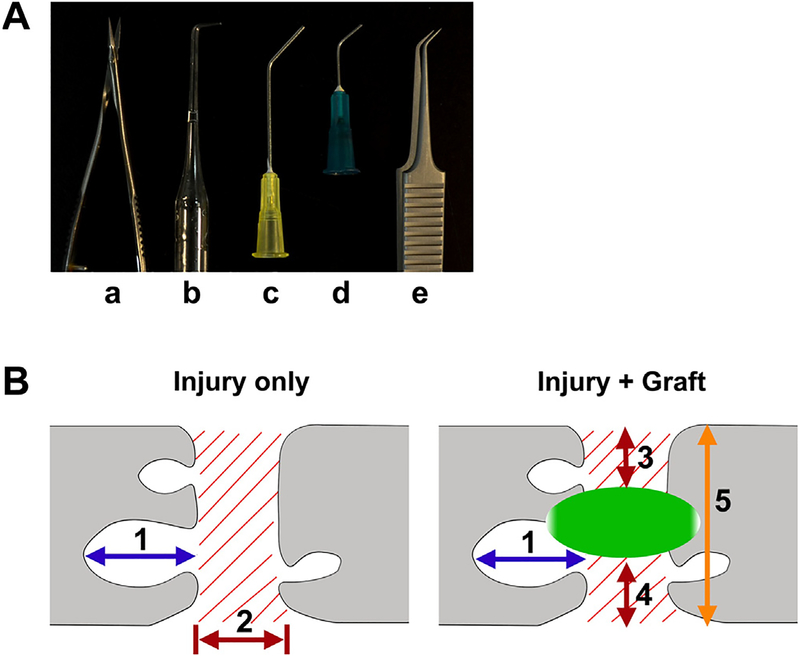
Fig. 1.
Surgical tools (A) to create a complete SCI include (a) iridectomy microscissors, (b–d) three self-made aspirators with different diameters in the tip, and (e) fine forceps for crush. Schematic diagrams (B) illustrate how the sizes of cavities (1) and fibrotic scars (2 or 3) were measured. The size of cavity is referred to the rostrocaudal distance (1) of the biggest one in each section. The rostro-caudal distance of fibrotic scarring (2) in lesion is defined in the spinal cord with injury only, while the vertical depth of fibrotic scars (3 + 4) is scaled and divided by the width of the section (5) in grafted cords.

