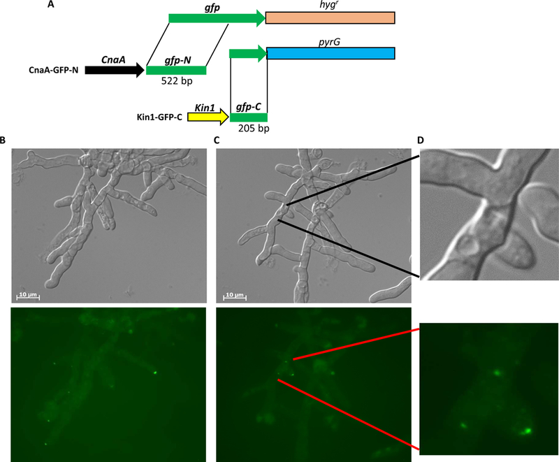
FIG. 3.

(A) Strategy for bimolecular fluorescence complementation. cnaA and kin1 are tagged to respective split form of gfp (gfp-N and gfp-C) with hygromycin resistance (hygr) and pyrG marker genes for selection. Both constructs were co-transformed into akuBKU80pyrG strain for expression and visualization of interaction. (B-D) BiFC microscopy showing interaction of Kin1-GFP-C and CnaA-GFP-N at tips and septa (indicated by arrowheads and arrows in lower panels) following caspofungin induced cell wall stress. Tip lysis seen in DIC images (upper panels; indicated by dotted arrows).
