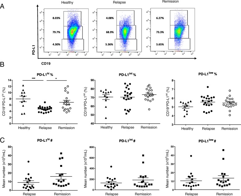
Fig. 2.
MS patients have deficiency of CD19+PD-L1hi cells during relapse. The percentage and the absolute number of CD19+PD-L1hi cells were measured in MS patients undergoing relapse (n = 20) and patients in remission (n = 17) and healthy controls (n = 11). Fresh PBMC was isolated from peripheral blood and surface stained for flow cytometry. a Representative flow-cytometry dot plot of PD-L1 and CD19 expression in total CD19+ B cells. b Scatter plots showing the percentage of CD19+PD-L1hi cells in MS-relapse, MS-remission, and HC. The frequency of CD19+PD-L1hi cells was significantly reduced in MS-relapse compared to MS-remission and HC (relapse vs remission: p = 0.0113, relapse vs healthy: p = 0.0007). All values show mean ± SEM. Data were analyzed by one-way analysis of variance (ANOVA) with Tukey’s multiple comparison post hoc analysis. *p < 0.05; ***p < 0.001. c Scatter plots showing the absolute number of CD19+PD-L1hi cells in MS-relapse and MS-remission. All values show mean ± SEM. Data were analyzed by unpaired t test. Ex vivo data were collected from peripheral blood samples taken during the time course of this study

