Figure 1.
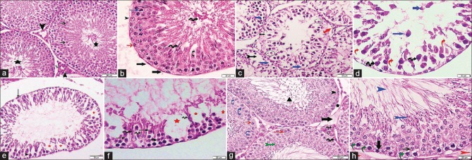
Photomicrographs of testicular sections stained with H and E. (a) Group I shows seminiferous tubules lined by several layers of rounded spermatogenic cells with central rounded nuclei. The spermatids are near the lumen (thin arrow) and the spermatozoa are in the lumen (stars). The interstitial Leydig cells (arrowheads) appear acidophilic with pale rounded nuclei and surround blood vessels (V) ×200. (b) A higher magnification of a seminiferous tubule surrounded by basal lamina and myoid cells (thick arrows). It displays spermatogonia (red arrow), primary spermatocytes (blue arrows), and spermatids (curved arrow) appear near to the lumen that contains spermatozoa (spiral arrows). Sertoli cell (arrowhead) with its vesicular nucleus can be seen ×400. (c) Group II reveals partial separation of the basement membrane (red arrow) with a lot of empty spaces (bifid black arrow). Some spermatocytes and spermatids have fragmented nuclei (circles) and others have deeply stained ones with vacuolated cytoplasm (blue arrows) ×200. (d) A higher magnification presenting loss of the normal architecture of spermatogenic cells, with the presence of empty spaces in between (red arrows). Some cells appear with deeply stained nuclei (spiral arrows) and some are shed off in the lumen (blue arrows) ×400. (e) Group III shows few layers of spermatogenic epithelium. Both spermatogonia (black arrow) and primary spermatocytes (arrowhead) have deeply stained nuclei. Empty spaces are still found within the tubule (red stars) ×200. (f) A higher magnification revealing spermatogenic cells with dark nuclei and deep acidophilic cytoplasm (black arrows). Loss of cellular connection between cells is apparent and there are empty spaces devoid of spermatogenic cells (red stars). Early sperms start to appear near the lumen (spiral arrows) ×400. (g) Group IV displays multiple layers of spermatogenic cells; spermatogonia (arrowhead) rest on basement membrane that is partially separated at some areas (star). There are primary spermatocytes (red arrows), spermatids near the lumen (bifid green arrow), spermatozoa inside it (►) and Sertoli cells (blue curved arrows). In interstitial tissue, Leydig cells with vesicular nucleus and prominent nucleolus (spiral arrow) surround blood vessels (V), with the presence of minimal exudate (thick black arrow) ×200. (h) A higher magnification displaying spermatogonia (thick arrow), primary spermatocytes (thin arrow), early sperms (bifid blue arrow), and mature sperms (blue arrowhead) within the lumen. Some cells are vacuolated (curved green arrows). There is slight exudate outside the tubule with separation of the basement membrane (red arrowheads) ×400

