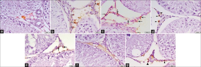
Figure 3.
Photomicrographs of testicular sections stained with CD34 immunohistochemical stain ×400. (a) Group I on day 5 displays no CD34 +ve HSCs. (b) Group II on day 5 shows some +ve CD34 cells with brown cytoplasmic reaction in the interstitial cells (arrows). (c) Group IV on day 5 reveals many CD34 +ve cells within the interstitial tissue (arrows). (d) Group I after 4 weeks shows positive immunoreactivity for CD34 in the walls of blood vessels in the interstitial tissue (arrowheads). No CD34 +ve cells can be detected. (e) Group II after 4 weeks presents few CD34 immunopositive HSCs in the interstitial tissue (arrows). (f) Group IV after 4 weeks has no CD34 immunopositive cells. (g) Group III after 6 weeks reveals few +ve HSCs (▲)

