Fig. 2.
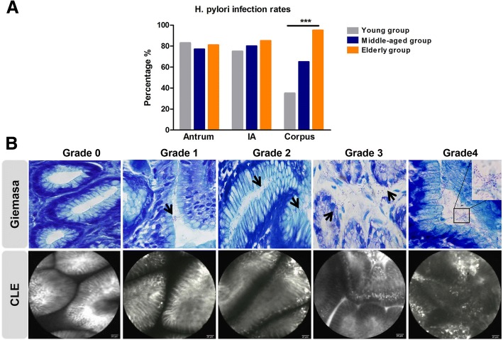
The distribution and severity of H. pylori in stomach in different age groups. a H. pylori infection rates in antrum, incisura angularis (IA) and corpus with increasing age. b The H. pylori colonization density was graded by Giemsa staining (upper panel) and confocal laser endomicroscopy (lower panel). A total of 20 high power (× 40 objective) microscopic fields were randomly choosed in each Giemsa staining sample, and the average scores of those 20 fields for each slide were defined as H. pylori density scores. *** p < 0.001

