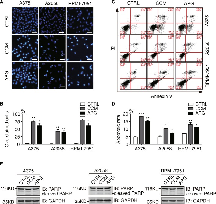
Fig. 2.
Apigenin and curcumin induce apoptosis in melanoma cells. a A375, A2058, and RPMI-7951 cells were treated with DMSO (control), curcumin (25 μM), or apigenin (30 μM) for 24 h before stained with Hoechst 33342. More than 200 cells were counted from 3 random views and percentages for apoptotic cells were shown in (b). Scale bar = 50 μm. c melanoma cells were stained with Annexin V and PI before flow cytometric analysis following 24 h treatment with DMSO, curcumin (25 μM), or apigenin (30 μM). d quantitation data of apoptosis from (c). e A375, A2058, and RPMI-7951 were treated as mentioned above and cleaved PARP was detected by Western blot analysis. GAPDH was shown as a loading control. All experiments were performed with 3 biological repeats. Error bars represent S.D. *P < 0.05, **P < 0.01, and ***P < 0.001

