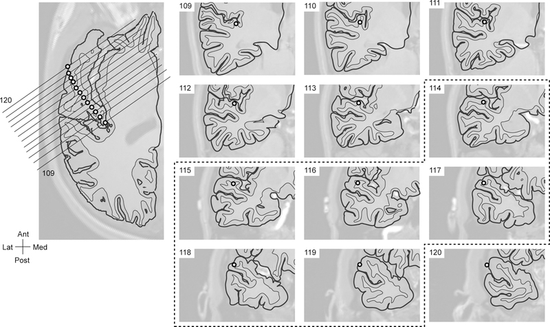
FIG. 4.

The location of each contact of the HG depth electrode. The location of each contact (numbered from 109 posteromedially to 120 anterolaterally) is shown on the axial image on the left and on the coronal images on the right. The electrode contacts that showed significant spikes during the seizure onsets were marked with a dashed line (from contact 114 medially through contact 119 laterally).
