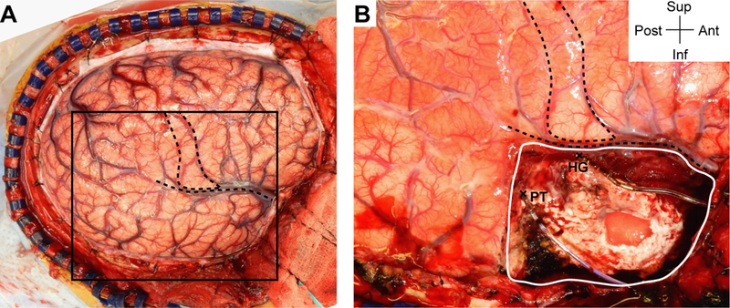
FIG. 5.

Intraoperative photograph demonstrating resection of seizure focus. A: Right frontotemporoparietal craniotomy (same image as Fig. 1A). The box corresponds to the approximate area shown in panel B. Gross anatomical landmarks (primary motor area and sylvian fissure) are indicated by dashed lines. B: An expanded view showing the extent of resection visible in this view (a solid white line) along with the HG depth electrode anteriorly and the PT depth electrode posteriorly that were kept in place during resection (entry points are marked by X symbols). Figure is available in color online only.
