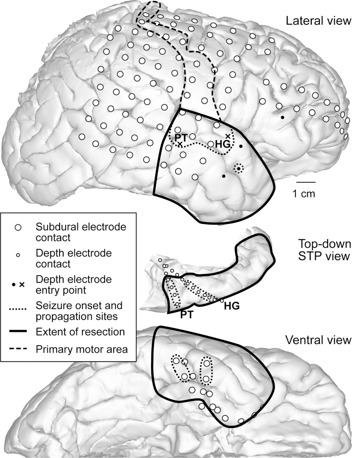
FIG. 6.

Reconstructed brain images with electrode contacts, showing the locations of the most significantly involved contacts during seizure onset and propagation, the extent of resection, and the HG and PT depth electrodes relative to the STP. The surface views were created by the FreeSurfer software based on preoperative and postoperative imaging studies. The lateral (upper panel) and ventral (lower panel) views show electrode contacts (large white circles), areas most significantly involved in seizure onset and propagation (dotted lines), the mapped primary motor area (dashed line), the extent of resection (solid line), and the entry points for the HG and PT depth electrodes (X symbols). The top-down view of the right STP (center panel) shows the locations of the HG and PT depth electrode contacts (small white circles).
