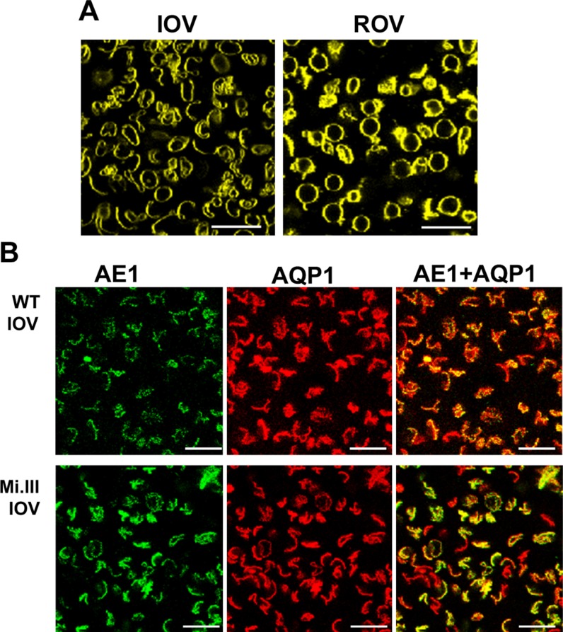
Figure 2.
Confocal imaging showed colocalization of AQP1 and band 3 on the surface of erythrocyte vesicles. A) Human RBC ghosts were resealed to form IOVs or ROVs. These vesicles were stained with the lipid-bound fluorophore Di-8-ANEPPS for visualization. B) Band 3 on the surface of IOVs was labeled with BRIC170-Alexa Fluor 488 (green fluorescence) and anti-AQP1-Alexa Fluor 568 (red fluorescence). Immunofluorescence-labeled IOVs from non-Mi.III (WT) and Mi.III RBC-IOVs showed substantial expressions of band 3 and AQP1. Various shades of orange and yellow fluorescence indicate different degrees of colocalization between band 3 and AQP1 on the surface of IOVs. Scale bars, 20 μm.

