
Fig. 1. Loss of p38α signaling correlates with enhanced tumorigenesis in human barrier tissues.
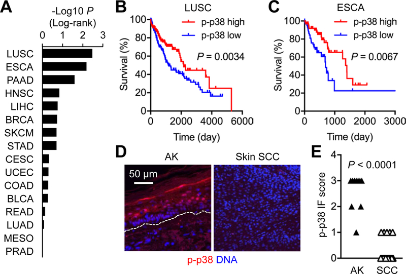
(A to C) Differential survival rates of the indicated TCGA patient groups with high and low phosphorylated (p-) p38 signals in their tumors are shown as P values by log-rank test. The cancer types are indicated by TCGA abbreviations, including LUSC (lung squamous cell carcinoma) and ESCA (esophageal carcinoma). The survival of LUSC and ESCA patients (n = 160 and 63 per group, respectively) is shown in Kaplan-Meier plots (B and C).
(D and E) Human actinic keratinosis (AK) skin and SCC tumor sections (n = 10 and 16, respectively) were analyzed by immunostaining (D). Dotted line, the epidermal-dermal boundary (D). p-p38 signals were graded from 0 to 3 based on immunofluorescence (IF) intensity per cell (E). P values by Mann-Whitney test.