
Fig. 2. Loss of p38α signaling results in enhanced skin tumorigenesis in mice.
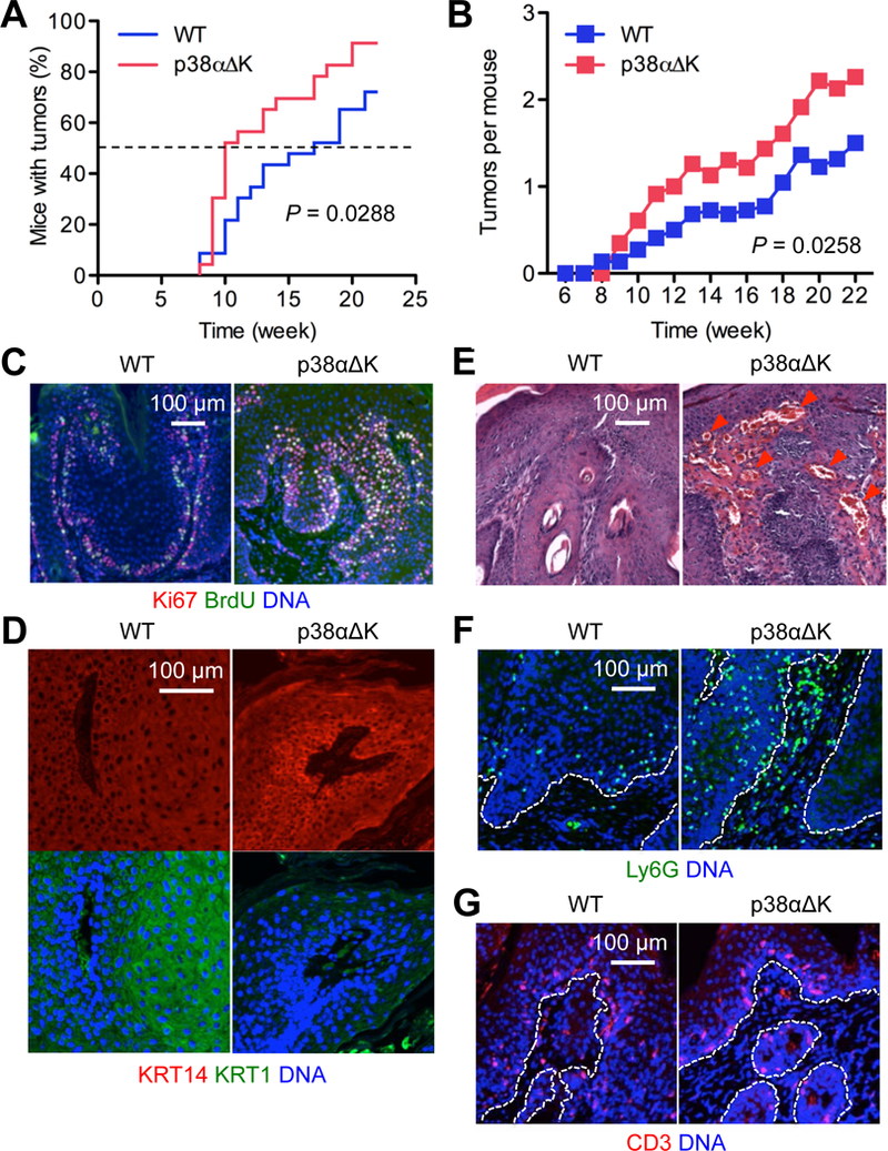
(A and B) Mice (n = 23) were subjected to DMBA-TPA skin tumorigenesis. Tumor incidence (A) and multiplicity (B) were determined over the indicated period. P values by log-rank test (A) and two-tailed unpaired Student’s t test (B).
(C to G) DMBA-TPA-induced tumor sections from mice were analyzed by immunostaining/counterstaining for the indicated molecules (C, D, F and G) and by H&E staining (E). Red arrowhead, peritumoral vasculature (E). Dotted line, the tumor-stroma boundary (F and G). Images are representative of three to five tissue sections.