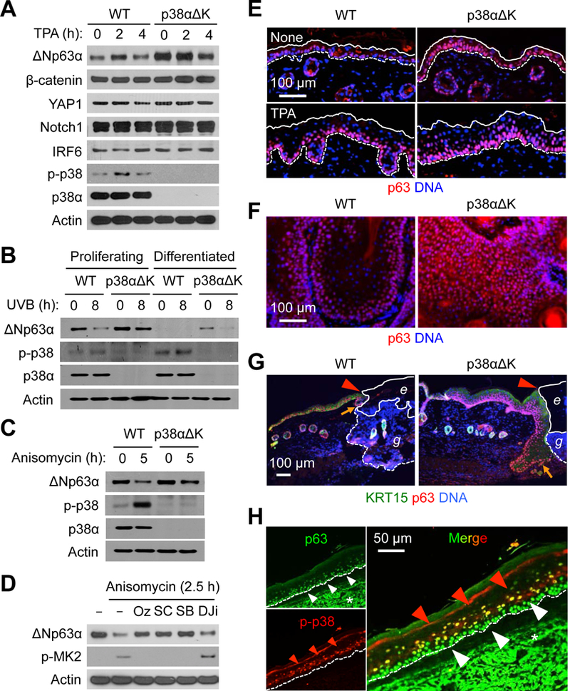
Fig. 5. p38α activity reduces p63 protein amounts and p63+ cell numbers in the epidermis and epidermal-derived tumors.

(A to D) Mouse (A-C) and human (D) keratinocytes were left unstimulated or stimulated with TPA (100 nM), UVB (75 mJ/cm2), and anisomycin (10 μg/ml). Whole cell lysates were prepared after the indicated durations of stimulation and analyzed by immunoblotting. p-, phosphorylated. Cells were preincubated with 5Z-7-oxozeaenol (Oz), SC409 (SC), SB202190 (SB), and D-JNKi (DJi) before stimulation (D). Blots are representative of three experiments.
(E to G) Steady-state (“None”) and TPA-treated skin sections (E), DMBA-TPA-induced tumor sections (F), and wound skin sections (G) from mice were analyzed by immunostaining/counterstaining for the indicated molecules. Solid and dotted line, epidermal margins and the epidermal-dermal boundary, respectively (E). Red arrowhead, wound margin; orange arrow, leading edge of regenerating epidermis; solid and dotted line, margins of eschar (e) and granulation tissue (g), respectively (G). Images are representative of five to seven tissue sections.
(H) Human AK skin sections were analyzed by immunostaining for the indicated molecules. Red and white arrowheads, epidermal cell layers with differential immunofluorescence signals; dotted line, the epidermal-dermal boundary; *, non-specific staining. Images are representative of three tissue sections.
