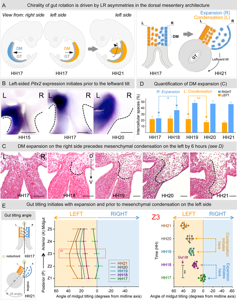
Figure 1: Expansion of the DM right side initiates gut rotation.

A The gut tube (GT, grey), in the chicken embryo at HH17 (mouse E9.5) suspended by the DM (blue-right; orange-left) undergoes a rotation to drive looping. This is driven by the leftward tilt (HH21, mouse E10.5) mediated by changes in cell architecture across the LR axis of the initially symmetric DM (HH17). B Pitx2 expression in the left DM appears prior to the initiation of the tilt (Pitx2 RNA ISH). C H&E staining of Z3 midgut sections from HH17–21 shows that right-sided expansion breaks the DM symmetry at HH18, prior to left-sided condensation at HH20. D Quantification of intercellular (cell-free) spaces from C; right expansion: p = 0.0001, left condensation: p = 0.0044, n = 10 embryos per stage. E Angle of midgut tilting from HH17–21, measured as displacement of GT from the midline (notochord) reveals significant increase in tilting at HH18 coincident with expansion (p = 0.0012, n = 10 embryos for HH17, HH18, n = 9 embryos for HH19, n = 7 embryos for HH20 and n = 6 embryos for HH21). Error bars represent mean ± SEM. See also Figure S1. Scale bars: B (100 µm); C (50 µm).
