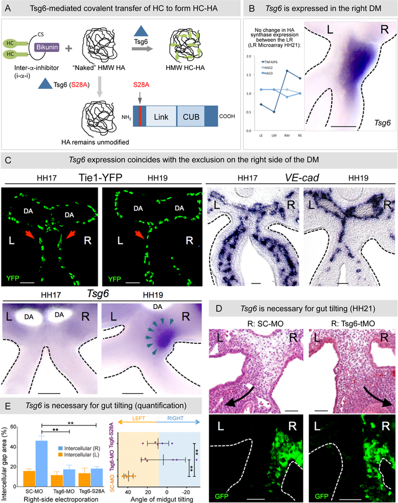
Figure 4: Tsg6 expression in the DM is right-sided and necessary for gut tilting.

A Tsg6 modification of HA via transfer of HC complexes. A catalytically inactive form of Tsg6 (S28A) fails to covalently modify HA. B Laser capture microarray and RNA ISH for Tsg6 in the chicken DM reveal right-sided expression at HH21. C Onset of Tsg6 expression in the right DM at HH19 (bottom panel) coincides with vascular exclusion (top panel left: Tie1-H2B-YFP quail embryos; right: RNA ISH for VE-cadherin). D Tsg6-MO-knockdown, pCAGEN-Tsg6 S28A overexpression causes reduced expansion and subsequent loss of tilting, quantified in E: Left panel: p = 0.0021 for SC-MO vs Tsg6-tMO, p = 0.0015 for SC-MO vs pCAGEN-Tsg6 S28A; Right panel: p = 0.0036 for SC-MO vs Tsg6-tMO, p = 0.0032 for SC-MO vs pCAGEN-Tsg6 S28A, n = 6/6 embryos for SC-MO, n = 6/6 embryos for Tsg6-tMO, n = 5/5 embryos for pCAGEN-Tsg6 S28A. Error bars represent mean ± SEM. See also Figures S2, S4, S5C, and S6B. Scale bars: B, C (100 µm); D (50 µm).
