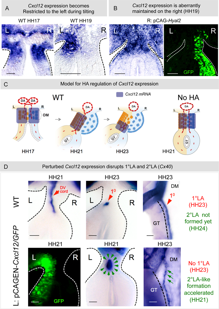
Figure 6: HA negatively regulates Cxcl12 mRNA expression.

A Bilateral Cxcl12 at HH17 in WT embryos becomes restricted to the left concurrent with expansion and vascular exclusion at HH19. B Depletion of HA from the right causes abnormal maintenance of Cxcl12 on the right at HH19 (p = 0.0108 for WT vs pCAGEN-Hyal2, n = 0/10 for WT, n = 6/10 for pCAGEN-Hyal2). C Model showing dynamics of Cxcl12 expression in WT embryos. Loss of HA aberrantly maintains Cxcl12 on the right and perturbs the DV gradient of Cxcl12 on the left. D pCAGEN-Cxcl12 in the left DM perturbs the normal 1°LA morphogenesis (red arrowhead: WT 1°LA formed from DV cords [red arrow]. Green arrows: L: pCAGEN-Cxcl12, GFP marks electroporated cells (left panel); middle panel: transverse slices: p = 0.0278 WT vs pCAGEN-Cxcl12, n = 0/5 slices for WT, n = 5/7 slices for pCAGEN-Cxcl12; right panel: whole embryo, p = 0.022 WT vs pCAGEN-Cxcl12, n = 0/10 embryos for WT, n = 3/5 embryos for pCAGEN-Cxcl12, RNA ISH for Cx40. Scale bars: A, B (100 µm); D (50 µm).
