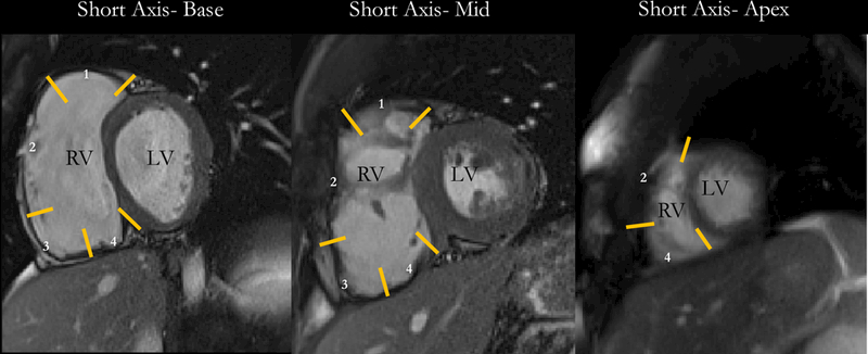
Figure 1-. Right Ventricular Segments.

Illustration of the segmentation of the RV on various short-axis levels. Endocardial and epicardial contours were drawn in a semi-automated manner from the anterior to the inferior RV insertion points. RV was segmented into outflow tract (1), free wall (2), angle (3), and inferior regions (4) in basal and mid-RV cuts and into free wall and inferior segments at the apex. RV= Right Ventricle; LV= Left Ventricle.
