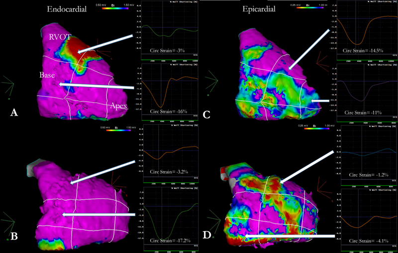
Figure 2-. Illustrative cases of the correlation between CMR-strain and EAM-scar.

Two representative cases of endocardial and epicardial voltage maps with corresponding regional strain analysis. Panels A and C correspond to a 25-year old female patient, mutation negative genotype, presenting with resuscitated sudden cardiac death with an RV ejection fraction of 36% and RV end-diastolic volume index of 115.2 ml/m2. Strain at the RVOT was severely depressed (−3.0%) but normal otherwise. On endocardial mapping, dense scar at the RVOT was found; epicardial mapping revealed slight drop in bipolar voltage but no scar. Panels B and D correspond to a 35-year old female patient, PKP2 positive genotype, presenting with symptomatic, non-resuscitated VT with an RV ejection fraction of 51% and RV end-diastolic volume index of 108.4 ml/m2. Strain analysis revealed depressed function at the angle, the RVOT and the mid-lateral wall. Although endocardial mapping revealed no voltage abnormalities, further epicardial mapping revealed large areas of dense scar at the corresponding anatomical segments (RVOT, angle and mid-RV). RV= Right Ventricle; RVOT= Right Ventricular Outflow Tract.
