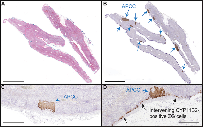
Figure 2. Representative images of APCC in IHA adrenals.

Representative images of hematoxylin and eosin (H&E, A) and CYP11B2 immunohistochemistry (B) are shown. All IHA cases harbored at least one APCC (blue arrows), supporting APCC as the common cause of IHA. Of the 15 adrenals studied, intervening zona glomerulosa (ZG) cells were negative (C) for CYP11B2 in 11/15 cases. Intervening ZG was diffusely positive (D, black arrow) for CYP11B2 only in 4/15 cases, arguing against diffuse aldosterone overproduction as the most common cause of IHA. Scale bars in A and B are 5mm, and in panels C and D are 1mm.
