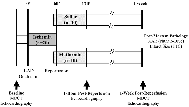
Figure 1: Experimental protocol.
All study personnel were blinded to treatment allocation until completion of data collection and analysis. Following baseline data collection including echocardiography and multi-detector computed tomography (MDCT) imaging, swine were subjected to a 60-minute occlusion of the left anterior descending (LAD) coronary artery in the closed-chest state. Contrast-enhanced MDCT was performed 5 minutes after the onset of LAD occlusion for assessment of the ischemic area-at-risk (AAR). Eight minutes prior to reperfusion, a bolus of metformin (5 mg/kg) or saline (vehicle) was administered via intravenous infusion in blinded fashion. Two minutes prior to reperfusion, an intracoronary infusion of metformin (1 mg/mL/min) or saline was initiated through the guide catheter engaged in the LAD and maintained until 15 minutes after reperfusion. Serial echocardiography was performed to assess LV function and MDCT imaging was repeated 1-week post-reperfusion to assess infarct size and LV ejection fraction. Immediately prior to euthanasia, the LAD was re-occluded, phthalocyanine blue was administered, and the heart was excised for post-mortem assessment of the ischemic AAR and infarct size. Please see text for additional details. LAD indicates left anterior descending coronary artery; MDCT, multi-detector computed tomography; AAR, area-at-risk; TTC, triphenyl tetrazolium chloride.

