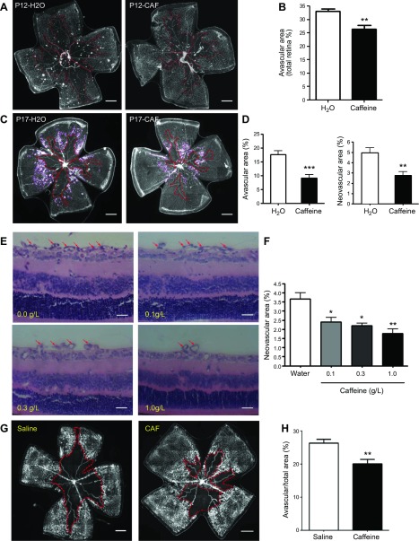
Figure 2.
Caffeine treatment reduces retinal vaso-obliteration at P12 and pathologic angiogenesis at P17 in OIR. Starting from the first day after delivery, pups were exposed to caffeine (Caf) via feeding nursing mothers with 0.1–1.0 g/L caffeine in drinking water. A, C, E) Whole-mount retinas (A, C) or retinal cross-section (E) from the water-OIR and caffeine-OIR groups were harvested on P12 (A) or -17 (C, E) and examined by immunostaining with isolectin B4, or histologic analysis of intravitreoretinal neovascular nuclei was performed. Representative images show avascular areas (indicated by red dotted line) at P12 (A) and -17 (C) and vitreoretinal neovascular tufts (indicated by purple line) in retinal whole-mounts at P17 (C). Scale bars, 500 μm. B, D) Areas of vaso-obliteration (B, D) and vitreoretinal neovascular tufts (D) were quantified and analyzed. **P < 0.01, ***P < 0.001; Student’s t test (n = 10/group). E, F) Representative images (E) show neovascular nuclei (red arrow) after treatment with caffeine at doses of 0–1.0 g/L in drinking water. Vitreoretinal neovascular tuft areas in whole-mount retinas were quantified and analyzed (F). Caffeine treatment dose-dependently reduced pathologic neovascularization. Scale bars, 20 μm. *P < 0.05, **P < 0.01; 1-way ANOVA, Bonferroni post hoc test (n = 10/group). G, H) Representative images (G) show areas of vaso-obliteration (indicated by red dotted line) after direct intraperitoneal injection of caffeine (10 mg/kg at P7 and 2.5 mg/kg P8–-16 daily) into pups. Quantitative analysis (H) shows that direct injection of the caffeine treatment reduced the avascular area compared with saline-treated pups. Scale bars, 500 μm. **P < 0.01; Student’s t test (n = 7–8/group).

