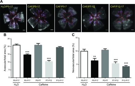
Figure 3.
Effective therapeutic windows for caffeine to confer protection against OIR. A) Pups were treated with water or caffeine (1 g/L) in different therapeutic windows (P0–17, P0–7, P7–12, and P12–17), and retinal vasculatures were analyzed by whole-mount isolectin B4 staining at P17 of OIR. Avascular areas are shown by red dotted line. Neovascularization tufts are indicated by purple line. Scale bar, 500 μm. B) Avascular area (%) was quantified as a percentage of the whole retinal surface (n = 7–9/group). C) Neovascularization tuft area (%) was quantified as a percentage of whole retinal area. **P < 0.01, ***P < 0.001; 1-way ANOVA, Bonferroni post hoc test (n = 7–9/group).

