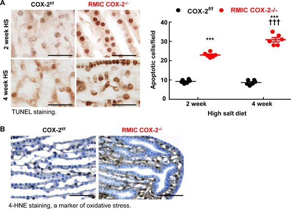
Figure 6. RMIC COX-2 deficiency led to apoptosis in the inner medullae/papillae in response to high salt diet.

A: RMIC COX-2−/− mice had increased papillary apoptotic cells as early as 2 weeks after a high salt diet, and more apoptotic cells were evident in both interstitial and epithelial cells 4 weeks after a high salt diet. ***P < 0.001 vs. corresponding COX-2f/f mice, †††P < 0.001 vs. 2 weeks of RMIC COX-2−/− mice; n = 6 in each group. Scale bar: 25 μM.
B: Four weeks after high salt intake, there was increased papillary oxidative stress in RMIC COX-2−/− mice, as indicated by 4-HNE-staining. Original magnification: x 160. All values are shown as mean ± SEM. P values were calculated by Student’s t test. Scale bar: 160 μM.
