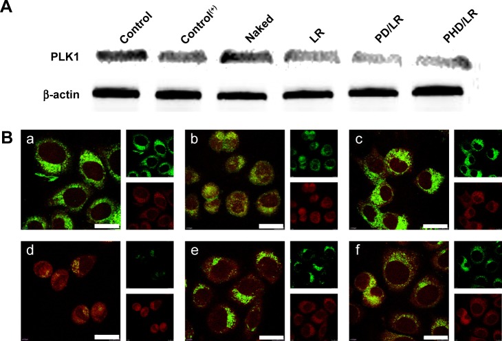
Figure 5.
In vitro PLK1 expression with Western immunoblotting (A) and immunofluorescence (B), respectively. Red and green fluorescence represents β-actin and PLK1, respectively. Scale bar =20 µm (a, b, c, d, e, and f represent Control, Control(+), naked siRNA, LR, PD/LR and PHD/LR, respectively).
Abbreviations: LR, lipoplex; PD, PHis-PSD; PEG, poly(ethylene glycol); PHD, PEG-PHis-PSD; PHis, poly(histidine); PSD, poly(sulfadimethoxine).

