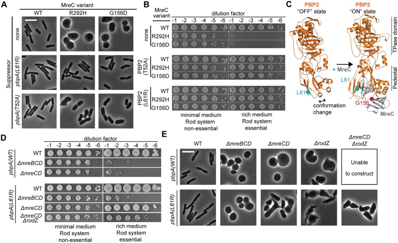
Fig 2. Substitutions in PBP2 suppress the growth and shape phenotypes of Rod system mutants.
A. Strains containing the indicated point mutations at the native genomic locus [PR164, PR165, PR166, PR127, PR128, PR129, PR131, PR124, PR125] were grown overnight in M9 CAA glu, diluted to OD600 = 0.05 in the same medium, and grown to exponential phase (OD600 = 0.2). Cells were gently pelleted, resuspended, and diluted in LB (OD600 = 0.025), then grown until the OD600 reached 0.2. At this time, cells were fixed, immobilized, and imaged using phase-contrast microscopy. All growth was at 30°C. Scale bar, 5 μm. B. Overnight cultures of the above strains were serially diluted and spotted on either M9 CAA glu agar (Rod non-essential) or LB agar (Rod essential). Plates were incubated at 30°C for either 40 h (M9) or 16 h (LB) prior to being photographed. C. Shown are E. coli PBP2 and PBP2-MreC structures modeled from PDB-5LP4 and PDB-5LP5 [27] using Phyre 2 [26]. PBP2 is orange with residue L61 in turquoise. MreC is gray with residue G156 in red. Structural information is lacking for the juxta-membrane region of PBP2 containing residue T52, and for the C-terminal domain of MreC containing residue R292 D. Strains containing the indicated mutations were grown and spotted as in (B) [Top to bottom: PR132, PR136, PR137, PR78, PR129, PR140, PR149]. E. The above strains were grown and prepared for phase-contrast microscopy as described in (A). Scale bar, 5 μm.

