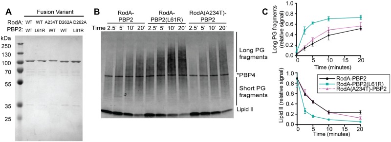
Fig 5. PBP2(L61R) stimulates glycosyltransferase activity of RodA.
A. Purified FLAG-RodA-PBP2 and mutant derivatives were subjected to SDS-PAGE and stained using Coomassie Blue. The molecular weight of the fusion proteins is approximately 114 kDa. B. Blot detecting the peptidoglycan product produced by the RodA-PBP2 fusion constructs incubated with extracted E. coli Lipid II for the indicated length of time. The product was detected by biotin-D-lysine (BDL) labeling with S. aureus PBP4. Note that labeled PBP4 protein appears as a band in the middle of the blot. C. The accumulation of long PG fragments and depletion of lipid II during three independent replicates of glycosyltransferase time-courses were quantified using densitometry. Error bars represent standard deviation.

