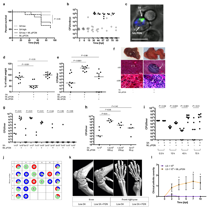
Figure 2. Gram-positive PGN augments S. aureus pathogenesis in animal models.
a, Survival curves of fish injected with low dose S. aureus SH1000 (150 CFU, SA low) and 5 ng polymeric M. luteus PGN (ML pPGN). S. aureus SH1000 high dose (1500 CFU, SA high) was injected as a positive control. Data are representative of three independent experiments; n ≥ 28, log-rank (Mantel-Cox) test. b, Growth of bacteria within embryos after co-injection with low dose S. aureus SH1000 (150 CFU) and 5 ng M. luteus PGN. Open circles, live and filled circles, dead embryos. n ≥ 60. c, In vivo imaging of Alexafluor 647 (blue) labelled M. luteus PGN (5 ng, ML PGN indicated by arrow) and S. aureus SH1000-GFP (150 CFU, SA indicated by arrow) 2 hpi. Within the zebrafish circulation valley, phagocytes were viewed at x 60 magnification). Images are representative of 5 embryos from two independent experiments. Scale bar 10 μm. d, e, BALB/c mice were injected i.v. with low dose (1x10⁶ CFU) S. aureus NEWHGkan with or without 500 μg M. luteus particulate PGN (pPGN) or soluble PGN (sPGN). Weight loss (d) and liver (e) CFU were measured. n = 10 per group; median value shown, Mann-Whitney two-sided test. f, Representative images of histopathological changes during infection. Arrows show 1, large abscess within liver parenchyma; 2, accumulation of extracellular S. aureus; 3, dense infiltrate of polymorphonuclear leukocytes (PMNs). Inset box at x10 magnification (scale bar 100 μm) is displayed at x100 (scale bar 10 μm) in bottom panels. n = 5 per group. g, Liver CFU recovered from BALB/c mice injected i.v. with a decreasing dose of S. aureus NEWHGkan with or without 500 μg M. luteus pPGN. n = 5 per group; median value shown, Mann-Whitney two-sided test. h, Liver CFU recovered from BALB/c mice injected i.v. with low dose (1x10⁶ CFU) S. aureus NEWHGkan with or without a decreasing dose of M. luteus pPGN. n = 5 per group; median value shown, Mann-Whitney two-sided test. i, Liver CFU at various time points after co-injection of low dose (1x10⁶ CFU) S. aureus with or without 500 μg M. luteus pPGN. n = 8 per group; median value shown, Mann-Whitney two-sided test. j, Livers from mice injected with low dose (1x10⁶ CFU; 1:1:1 mixture of NewHG EryR, TetR or KanR, n = 5 per group S. aureus NEWHGkan plus 500 μg M. luteus PGN were harvested. Individual abscesses were dissected and bacterial CFU enumeration from each abscess was determined (A1-A4). Bacterial CFUs from residual liver tissue post dissection (-) was also enumerated and added to the abscess CFUs to provide a total CFU count for each liver (L). k, Micro-CT imaging of knee and front right paw of an NMRI mouse injected i.v. with low dose (1x10⁶ CFU) S. aureus LS-1 with or without 1 mg M. luteus pPGN. Images are representative of 10 animals. l, Clinical arthritis severity of NMRI mice injected i.v. with S. aureus LS-1 low dose (1x10⁶ CFU) and 1 mg M. luteus pPGN. dpi, days post infection. n = 10 per group; error bars, mean and s.e.m, Mann-Whitney two-sided test. * P < 0.05.

