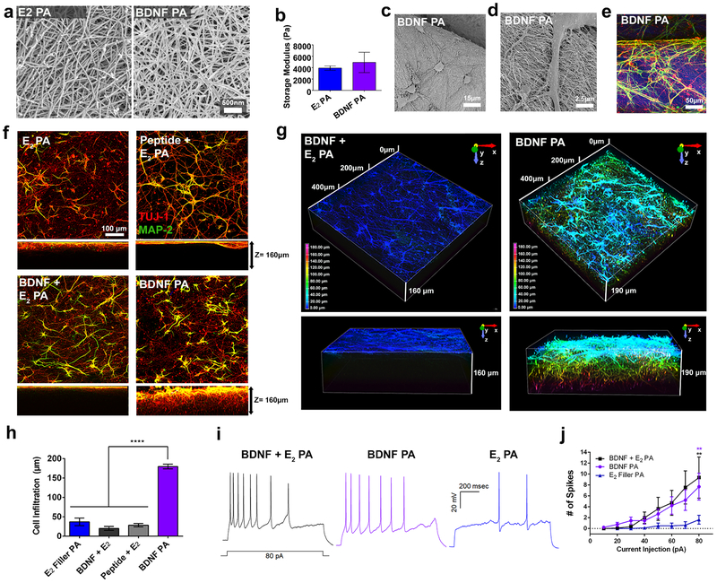
Figure 5. Infiltration and maturation of cortical neurons in BDNF PA scaffolds.
(a) SEM showing nanofibers in E2 PA and BDNF PA gels. (b) Storage moduli of E2 PA and BDNF PA. (c-d) Scanning electron micrograph and (e) confocal micrograph images showing cortical neurons seeded on top of 3D PA scaffolds for 1 week in vitro. (f) Confocal images showing top and side (z-stack =160 μm) view sections of cells cultured on PA gel scaffolds for 1 week in vitro (images show cells stained with MAP-2 (dendritic marker, green), and Tuj-1 (neuronal marker, red). (g) Depth-coded z-stack reconstructions showing cell infiltration after 1 week in vitro. (h) Quantification of cell infiltration depth in E2 PA, BDNF + E2 PA, BDNF peptide + E2 PA, and BDNF PA gels. (i) Representative traces of action potentials elicited by 1 second, 10pA current injection steps from 10–80pA recorded from neurons in BDNF + E2 PA, BDNF PA and E2 PA gels, 1 week in vitro. (j) Plot of current injection versus number of spikes for conditions in (i). **P<0.01 and ****P<0.0001. LSD test (b and h) and ANOVA followed by posthoc analysis was used for current clamp recordings (j).

