
Fig. 3. Cancer type-specific modeling on-chip.
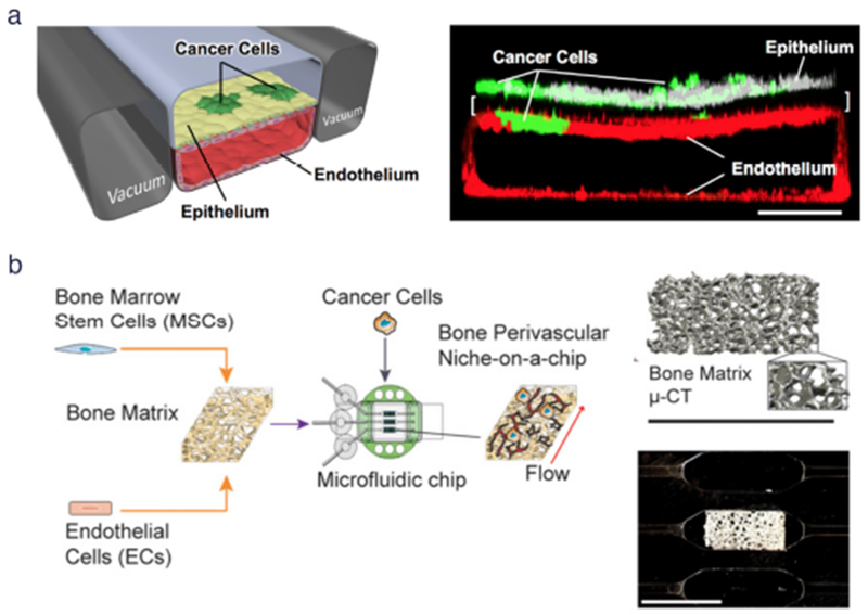
(a) (Left) Schematic diagram of a cross-section through 2-channel microfluidic lung-on-a-chip device. (Right) Confocal fluorescence micrograph of a cross-section of the two central cell-lined channels of an alveolus chip. NSCLC cells are labeled with GFP and endothelium with RFP, as shown. Reproduced from Hassell et al46 with permission from Cell Press, (b) (Left) Workflow for generating bone perivascular (BoPV) niche for studies of breast cancer colonization. (Right Top) Bone tissue reconstruction based on micro-computed tomography (μ-CT) data. (Right Bottom) Rectangular-shaped bone matrix in microfluidic chip. Reproduced from Marturano-Kruik et al47 with permission from the National Academy of Sciences.