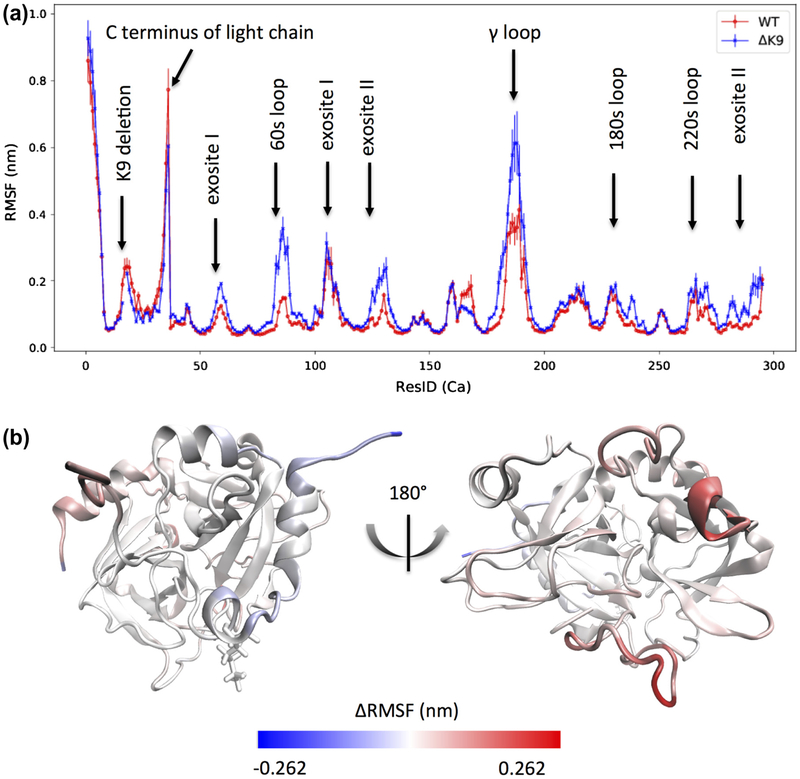
Figure 3.
Root-mean-square fluctuations (RMSF) for the alpha carbons of ΔK9 (Mutant) and wild-type (WT) thrombin. The blue and red colors in (a) depict the ΔK9 mutant and wild-type thrombin respectively. The residue indexes in (a) follow our sequential residue numbering scheme that all residues in the light and heavy chains are numbered from 1 to 295 and the LYS9 in the light chain thereby has a residue ID of 17. The known sites with distinct RMSF are indicated by labels. The thrombin molecule is colored based on the subtractions of RMSF (Mutant-WT) in (b) to indicate the location of the significantly affected regions.

