
Fig. 3.
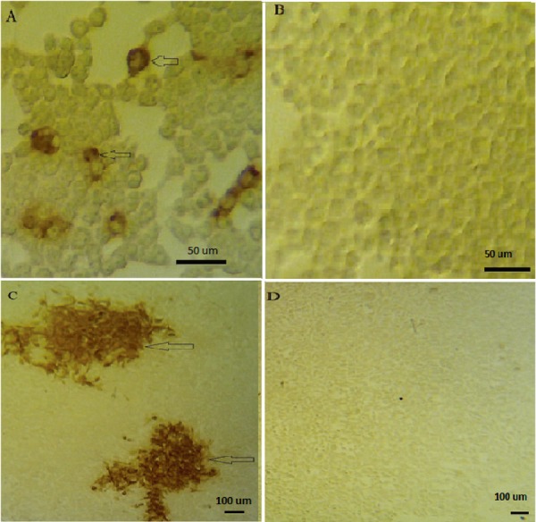
Detection of rabies virus antigen by immunocytochemistry assay to visualize the infected and uninfected cells following titration procedures. Quantitative assay to determine TCID50 (A and B) and ICC to determine focus forming units (FFU) (C and D). The infected and uninfected cells are shown, respectively, as cells with dark brown cells (arrow), and unstained cytoplasm.