
Figure 7.
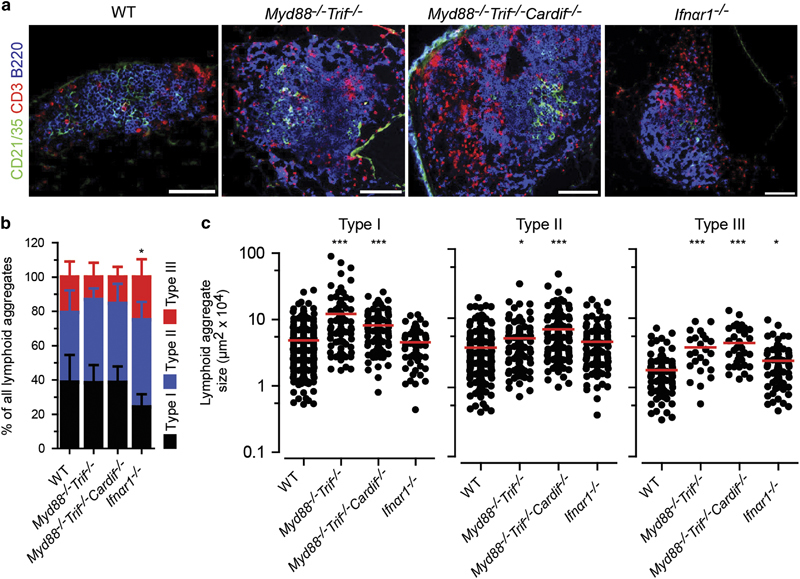
Lack of MyD88, TRIF, Cardif and IFNαR1 signaling does not affect the development of E. coli-induced BALT. (a) Representative immunofluorescence micrographs from lung sections taken from day 18 following E. coli infection in WT, Myd88 −/− Trif −/−, Myd88 −/− Trif −/− Cardif −/− and Ifnαr1 −/− mice. (b) Relative frequency and (c) size of type I, type II and type III lymphoid aggregates on day 18 following E. coli infection. Data (b, c) collected from seven WT, 2 Myd88 −/− Trif −/−, 3 Myd88 −/− Trif −/− Cardif −/− and 3 Ifnαr1 −/− mice, in which four lung sections were analyzed from at least 2 independent experiments. Error bars (b) represent means±s.d.; bars (c) represent median values; *P<0.5; ***P<0.001. The color of stars (b) relates to the type of aggregate, as indicated. Scale bars=100 μm.