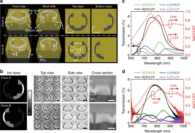
Fig. 1. Unit-cell design.
a Schematic of the chiral stepped nanoaperture in its two enantiomeric forms (Form A and Form B). The thickness of the gold film is H = 180 nm. The top and bottom layers of the aperture have thicknesses of h1 = 80 nm and h2 = 100 nm, with the cross-sections of the two layers illustrated. The dimension parameters are indicated as p = 360 nm, r = 140 nm, b = 120 nm, α = 60°, g1 = 20 nm, and g2 = 40 nm. Incoming waves are illuminated vertically onto the top layer and transmitted out of the bottom layer. b Normalized ion-dose distributions and SEM images of the stepped nanoapertures fabricated using the grayscale focused ion-beam milling method. Sideview images are captured with a visual angle of 52° to the surface normal. Scale bar: 100 nm. c Simulated and d measured transmission spectra of the stepped nanoapertures in Form A for different incident/output handedness combinations together with the corresponding CDT and CPR spectra

