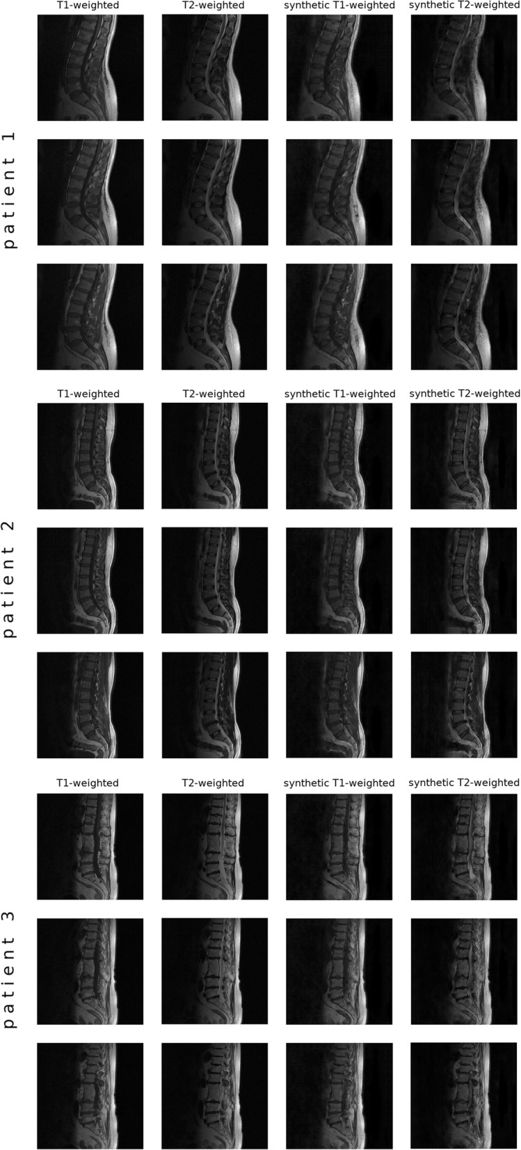
Fig. 4.

Representative results (three slices for three different patients) of the translation from T1W to T2W MRI and vice versa. In the first patient, Type II Modic changes were correctly represented in both synthetic images, whereas for the Type I change in the third patient the synthetic T2W image showed a low signal instead of a high one. The L4-L5 disc protrusion of the second patient was accurately represented in the synthetic T2W image
