FIGURE 9.
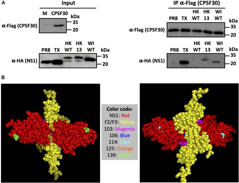
Amino acid changes L103F, I106IM, P114S, G125D and N139D in H9N2 NS1 are required for interaction with CPSF30. (A) Analysis of NS1-CPFS30 interaction by co-immunoprecipitation. CFSF30 was expressed in HEK293T cells, mixed with the indicated NS1 proteins synthetized in vitro and immunoprecipited using an anti-FLAG resin. Input (left) and immunoprecipitated (right) proteins were detected by Western blot using antibodies specific against the FLAG (CPSF30) or HA (NS1) epitope tags. Molecular mass markers (in kDa) are indicated. (B) Tridimensional structure of the NS1 effector domain binding to F2/F3 region of CPSF30: Monomers of IAV NS1 protein are represented in red. The F2/F3 region in CPSF30 involved in interaction with NS1 is represented in yellow. NS1 residues 103, 106, 114, 125, and 139 involved in interaction with CPSF30 are showed in different colors in the figure legend. The model structure was generated using Cn3D and is based on the NS1 of A/Udorn/72 H3N2 (PDB entry 2RHK).

