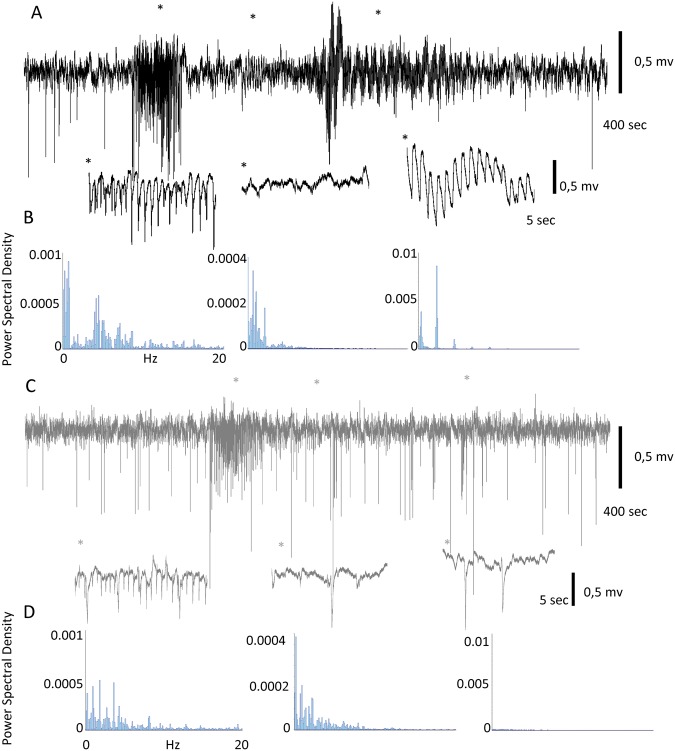
Figure 6.
Magnet reduces sensory evoked seizures in the primate. (A) Visual evoked seizure recorded in V1 under control conditions. Areas marked with * are expanded below. (B) Power spectrum density analysis of the 3 epileptiform elements expanded in A (note the different scales on the Y axis). (C) Recording of the longest seizure evoked in the presence of the magnet (high-frequency stimulation was necessary to evoke it). In this case only, the initial paroxysmal activity (with a lower amplitude) is present. (D) Power spectrum density analysis of the 3 elements expanded in C (same scale as in A is used to enable direct comparisons).

