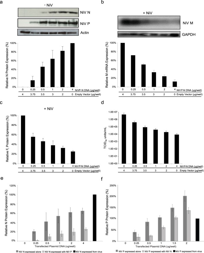
Figure 5.
Effects of increasing recombinant NiV N proteins co-expressed with NiV P proteins on NiV replication. Cells were transfected with increasing amounts of plasmid DNA, containing both the NiV N gene and NiV P gene, which are driven by individual promoters encoded on one construct for 48 hours. (a) Cells were harvested and analyzed by western blot using a monoclonal antibody against NiV N. Following transfection, a parallel set of cells were infected with NiV at an MOI of 1 for 24 hours. (b) RNA was extracted from NiV infected cells and analyzed by northern blot using a probe designed against the M gene. Northern blots were quantified by densitometry and normalized to GAPDH. (c) Total protein was harvested from NiV infected cells and analyzed by western blot for the expression of NiV F0 proteins. (d) Supernatants were harvested, viral loads were determined by endpoint titration, and TCID50/ml was calculated. (e) Cells were transfected with increasing amounts of plasmid DNA expressing NiV N and/or NiV P protein for 48 hours or infected with NiV at an MOI of 1 for 24 hours. Total protein was harvested and a western blot was performed to visualize the presence of NiV N proteins or (f) NiV P proteins. All western blots were quantified by densitometry and normalized to actin. All experiments were carried out in triplicate and standard deviations of the mean were calculated. Blots have been cropped to ease visualization.

