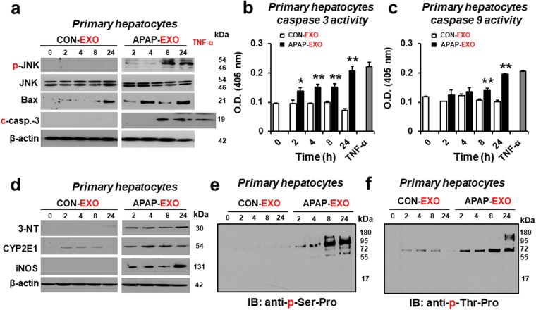
Figure 6.
Time-dependent increases in apoptosis-related, nitroxidative stress marker and phosphorylated proteins following exposure to APAP-derived exosomes. (a) Immunoblot showing relative amounts of p-JNK/JNK, Bax, cleaved caspase-3 and β-actin, a loading control (n = 4/sample). (b and b) Caspase 3 and 9 activities (n = 8/sample). (d) Immunoblot showing relative amounts of nitroxidative stress markers, 3-NT, CYP2E1, and iNOS compared to β-actin and (e,f) phosphoproteins containing p-serine-proline (e) or p-threonine-proline (f) in primary hepatocytes treated with APAP-derived or CON-derived exosomes for 0, 2, 4, 8, and 24 h, as indicated (n = 4/sample). Full-length immunoblots are presented in Supplementary Figure 7.

