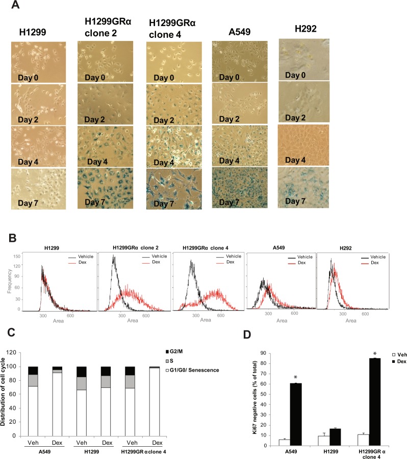
Figure 4.
Induction of senescence phenotype in lung adenocarcinoma model cell lines by extended Dex treatment. H292, A549, H1299, H1299GR Clone 2 and H1299GR Clone 4 cells were treated with Dex (100 nM) for the indicated periods and the cells were stained to assess expression of β- galactosidase (blue staining) (Panel A). The same cell lines were treated with either vehicle or Dex (100 nM) for 4 days (H1299GR Clone 2 and H1299GR Clone 4) or for 7 days (H1299, H292, A549) and the distribution of relative cell size was determined by measuring the area of individual cells using the Amnis Mark II imaging cytometer (Panel B). A549, H1299 parental, and H1299GR clone 4 cells were plated at 20 percent confluence; the cells were then treated with either vehicle or Dex (100 nM) for 7 days, replenishing the treatments every 3 days. Cells were then analyzed by flow cytometry to measure cell cycle distribution (Panel C) and Ki67 negativity (Panel D). *P, 0.0004.

