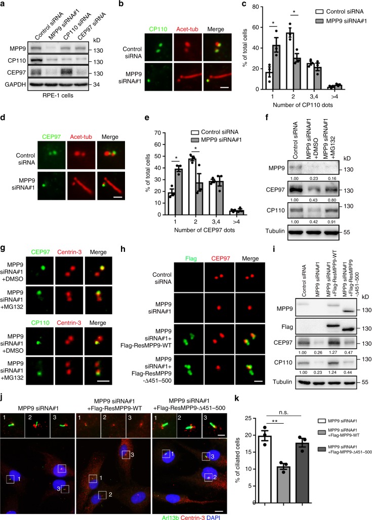
Fig. 4.
MPP9 recruits CEP97 and CP110 at the distal end of the mother centriole in hTERT RPE-1 cells. a Immunoblots showing depletion of MPP9, CP110, or CEP97 by siRNA in hTERT RPE-1 cells. GAPDH was used as a loading control. b Immunostaining of acetylated-tubulin (Acet-Tub, red) and CP110 (green) in the control- or MPP9-siRNA-treated hTERT RPE-1 cells. c Quantification of the percentage of cells with the indicated number of CP110 dots from b. d Immunostaining of Acet-Tub (red) and CEP97 (green) in the control- or MPP9-siRNA-treated hTERT RPE-1 cells. e Quantification of the percentage of cells with the indicated number of CEP97 dots from d. f Immunoblots showing expression of the indicated proteins in MPP9-depleted hTERT RPE-1 cells after treatment with DMSO or MG132 for 4 h. Tubulin was used as a loading control. Relative amounts of MPP9, CEP97, and CP110 were quantified and normalized to tubulin. g Immunostaining of Centrin-3 (red) and CEP97 (green, upper) or CP110 (green, lower) after treating with DMSO or MG132 for 4 h in MPP9-depleted hTERT RPE-1 cells. h Immunostaining of CEP97 (red) and Flag-siRNA-resistant MPP9 (Flag-ResMPP9, green) or Flag-ResMPP9-△451-500 (green) in MPP9-depleted hTERT RPE-1 cells. i Immunoblots showing expression of the indicated proteins in hTERT RPE-1 cells transfected with MPP9-siRNA and Flag-siRNA-resistant MPP9 wild-type (Flag-ResMPP9-WT) or lacking 451–500 aa mutant (Flag-ResMPP9-△451–500). Tubulin was used as a loading control. Relative amounts of CEP97 and CP110 were quantified and normalized to tubulin. j Immunostaining of Centrin-3 (red) and Arl13B (green) after transfection of Flag-ResMPP9-WT or Flag-ResMPP9-△451–500 in MPP9-depleted hTERT RPE-1 cells. DNA was stained with DAPI (blue). k Quantification of ciliogenesis in MPP9-depleted hTERT RPE-1 cells overexpressing Flag-ResMPP9-WT or Flag-ResMPP9-△451–500. For c, e, k, bars represent the means ± S.E.M for three independent experiments. n.s., not significant, *p < 0.05, **p < 0.01, as determined by unpaired two-tailed Student’s t-test (c, e), and one-way ANOVA analysis (k). Scale bars: 5 μm (j main); 1 μm (b, d, g, h, j insets)

