Fig. 5.
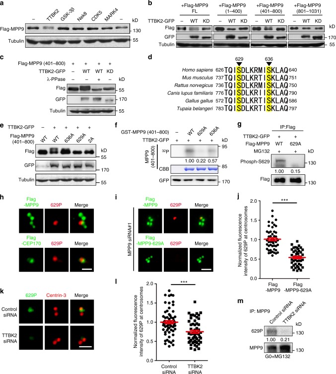
TTBK2 phosphorylates MPP9 mainly at S629 site. a Immunoblots of lysates from HEK293T cells co-overexpressing Flag-MPP9 and the indicated cilia-related kinase. Tubulin was used as a loading control. b Immunoblots of lysates from HEK293T cells co-overexpressing Flag-tagged MPP9 full-length (FL) or the indicated MPP9 truncation mutants with TTBK2-GFP-WT or TTBK2-GFP-KD. Tubulin was used as a loading control. WT, wild-type; KD, kinase-dead. c Immunoblots of lysates from HEK293T cells co-overexpressing Flag-tagged MPP9 (401–800 aa) and TTBK2-GFP-WT or TTBK2-GFP-KD after treatment with λ-PPase. Tubulin was used as a loading control. d Mass spectrometric analysis showing phosphorylation sites of MPP9 at S629 and S636. e Immunoblots of lysates from HEK293T cells co-overexpressing TTBK2-GFP and Flag-MPP9 (401–800 aa)-WT, or the indicated unphosphorylatable mutants. Tubulin was used as a loading control. f GST-MPP9 (401–800 aa)-WT, or the indicated mutants were subjected to a TTBK2 kinase assay in vitro followed by autoradiography. Coomassie blue (CBB) staining showed GST-tagged proteins. Relative amounts of phosphorylated MPP9 signals were quantified. g Lysates of HEK293T cells co-overexpressing Flag-tagged MPP9-WT or the S629A mutant and TTBK2-GFP were subjected to immunoprecipitation (IP) and immunoblotting with anti-Flag and anti-Phospho-S629 antibodies. Relative amounts of S629-phosphorylated MPP9 were quantified. h Immunostaining of S629-phosphorylated MPP9 (red) and Flag in Flag-MPP9 (green, upper) or Flag-CEP170 (green, lower) overexpressing hTERT RPE-1 cells. i Immunostaining of S629-phosphorylated MPP9 (red) and Flag in Flag-MPP9 (green) or Flag-MPP9–629A (green) overexpressing hTERT RPE-1 cells after depleting MPP9. j Quantifications of S629-phosphorylated MPP9 signals in i (n = 50 cells for each group). k Immunostaining of S629-phosphorylated MPP9 (green) and Centrin-3 (red) in control- or TTBK2-siRNA transfected hTERT RPE-1 cells. l Quantifications of S629-phosphorylated MPP9 signals in k (n = 50 cells for each group). m Lysates of control-siRNA or TTBK2-siRNA transfected hTERT RPE-1 cells after serum starvation were subjected to immunoprecipitation and immunoblotting with the indicated antibodies. Relative amounts of S629-phosphorylated MPP9 were quantified and normalized to MPP9. For j, l, bars represent the means ± S.E.M for three independent experiments. ***p < 0.001, as determined by unpaired two-tailed Student’s t-test. Scale bars: 1 μm (h, i, k)

