
Fig. 6.
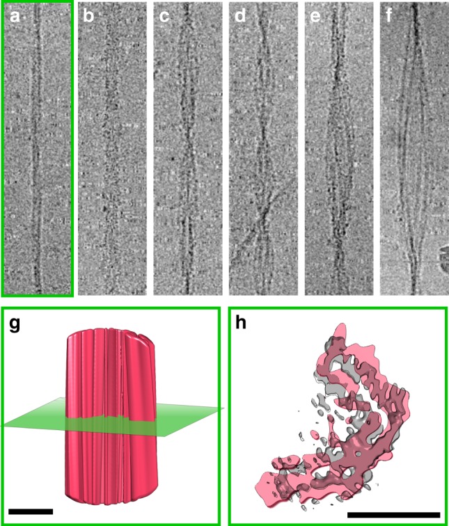
Variety in fibril morphology. Six different morphologies were identified in the non-entangled fibrils constituting the cryo-EM dataset. These range in size from apparent single protofilaments (a), through a series of apparent two protofilament fibrils (b–d), the structure of one of which has been described here at 3.9 Å resolution (panel c), to assemblies of multiple protofilaments (e, f). Each panel (a–f) is 500 Å wide. Side view (g) and cross-section through (h) a low-resolution reconstruction of morphology (a) showing density consistent with an L-shaped subunit. Each scale bar in panels (g) and (h) is 50 Å in length