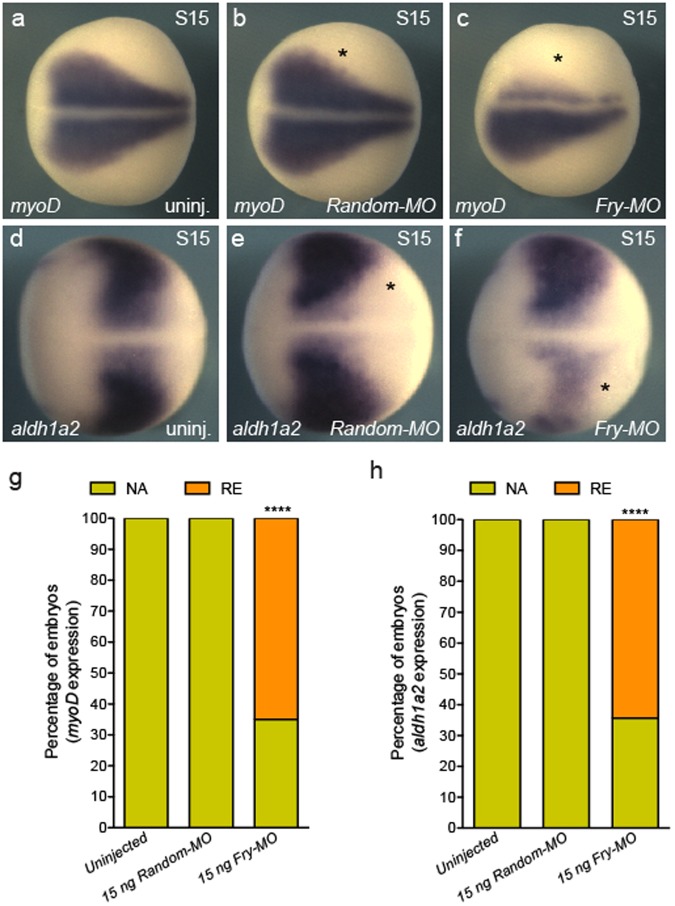
Figure 4.
Expression of paraxial mesoderm marker genes is reduced in Fry-depleted embryos. (a–c) In situ hybridization of S15 (stage 15) embryos for myoD. (g) Percentage of embryos with reduced (RE) or not affected (NA) myoD expression field revealed by in situ hybridization. Uninjected (N = 3, 92), Random-MO (N = 2, 36), Fry-MO (N = 4, 85). (d–f) In situ hybridization of S15 embryos for aldh1a2. (h) Percentage of embryos with reduced (RE) or not affected (NA) aldh1a2 expression field revealed by in situ hybridization. Uninjected (N = 3, 75), Random-MO (N = 2, 36), Fry-MO (N = 3, 76) (N = number of experiments, number of embryos). As indicated on each panel, 8-cell embryos were injected 1x V2 with 15 ng of the MOs or left uninjected. Asterisk indicates the injected side of the embryo. Representative embryos are shown. Data in graphs is presented as means. Statistical significance was evaluated using Fisher’s exact test ***p < 0.0001.

