Figure 4.

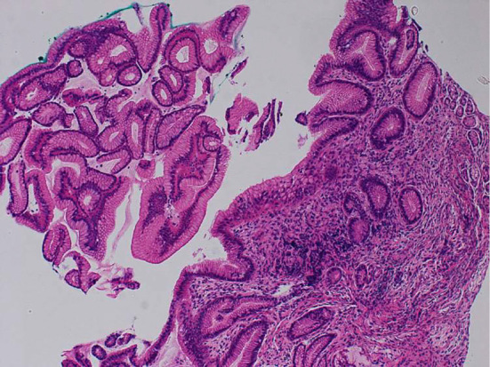
Pathological images of the biopsy specimens. A gastric biopsy showing regenerative changes, little inflammatory infiltration, and no abnormal findings in the gastric mucosal epithelium (Hematoxylin and Eosin staining, ×20).

Pathological images of the biopsy specimens. A gastric biopsy showing regenerative changes, little inflammatory infiltration, and no abnormal findings in the gastric mucosal epithelium (Hematoxylin and Eosin staining, ×20).