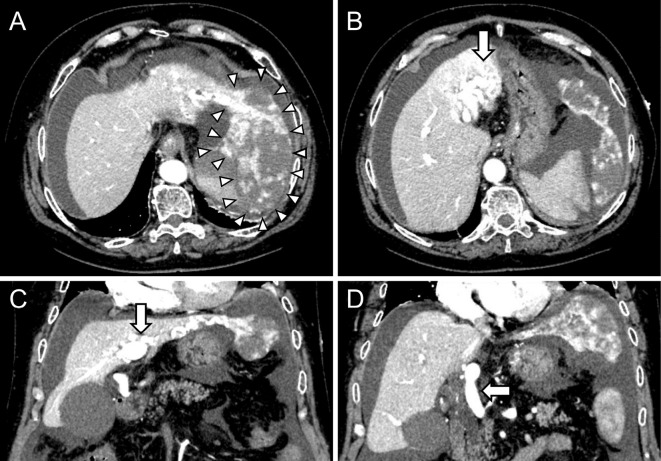
Figure 1.
Dynamic CT images showed a 10-cm pedunculated giant hepatic mass suggesting hemangioma in the left lobe of the liver (arrowheads) with massive ascites (A), an enlarged and tortuous hepatic artery (arrow) (B), an AP shunt (arrow) (C), and retrograde enhancement into the trunk of the portal vein (arrow) (D). AP: arterio-portal

